 D
D
This talk reports the results of work by many people, including Nicky Cullum, Principal Investigator, Jo Dumville, Trial Coordinator, Andrea Nelson, Co-Investigator, David J Torgerson, Head of York Trials Unit, Martin Bland, Senior Trial Statistician, Gill Worthy, Trial Statistician, Cynthia Iglesias, Senior Health Economist , Marta Soares, Health Economist, Christopher Dowson, Senior Microbiologist, Joanne L Mitchell, Microbiologist, and many more on the VenUS II team.
Larval therapy has been claimed to remove slough and necrotic tissue, promote wound healing and reduce wound bacteria including MRSA.
This idea is not just found among the healthcare professions. I came across this in one of Bernard Cornwell’s Peninsular War novels:
. . . ‘You’re going to be as right as rain, Jorge,’ Sharpe said, ‘so long as we keep that wound clean.’ He glanced at Harper. ‘Maggots?’
‘Not now, sir,’ Harper said, ‘only if the wound goes bad.’
‘Maggots?’ Vincente asked faintly. ‘Did you say maggots?’
‘Nothing better sir,’ Harper said enthusiastically. ‘Best thing for a dirty wound . . . I always carry a half-dozen.’
(Bernard Cornwell, Sharpe’s Escape, page 280, Harper Collins, 2004.
We tested these claims in people with sloughy and/or necrotic venous leg ulcers.
While this study was being planned, there was a development in larval technology. Bagged larvae were introduced, where the maggots were enclosed in mesh bag like a tea bag. It was hoped that this would make larval therapy more acceptable to both nurses and patients. We therefore included bagged larvae as another treatment, in addition to the standard loose larvae.
There were two objectives to the VenUS II trial:
VenUS II was a pragmatic, three-armed randomised controlled trial comparing hydrogel, loose larvae and bagged larvae. Patients with venous or mixed aetiology ulcers with at least 25% coverage of slough and/or necrotic tissue were eligible for the trial.
Participants were randomised to one of three groups:
These were all intended as debridement treatments, to remove the slough and necrotic tissue, so these treatments were applied only during debridement.
We intended to recruit 200 participants in each group, a total of 600. This would give us 89% power to detect a reduction in median healing time from 20 weeks to 14 weeks, allowing for 15% attrition.
Allocation was stratified for:
The primary outcome variable was to be time to complete healing of the largest eligible ulcer, with follow-up for one year.
The secondary clinical outcomes included:
All analyses would be adjusted for area of ulcer, duration, ulcer type, and trial centre.
Exactly how were we going to measure the primary outcome, the time to complete healing of the reference ulcer? The reference ulcer was defined as the largest ulcer containing at least 25% slough or necrotic tissue and was the ulcer receiving the trial treatment. It was decided that two assessors, blind to treatment group, would independently assess photographs of ulcers considered to be healed by the research nurses. They would agree on the point at which the ulcer healed. In the event, there were problems with this, as sometimes photographs were not available or not clear. In that case, the date as recorded by the research nurse on the ulcer healing form was used.
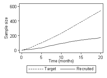
There were many delays and recruitment was much poorer than anticipated.
Figure 1 shows a typical graph reporting progress in recruitment.
We considered possible extensions to the trial, calculating the estimated recruitment for different extension periods:
| 6 months | 170 | |
| 12 months | 260 | |
| 18 months | 370 | |
| 24 months | 480 | |
| 30 months | 600 |
We decided to ask for and we obtained an extension of 18 months, to recruit 370 participants.
We failed!
267 participants were finally recruited from 18 centres.
| loose larvae: | 94 | |
| bagged larvae: | 86 | |
| hydrogel: | 86 |
Figure 2 shows the proportion healed at different times for all participants in VenUS II.
The graph shows the proportion estimated to have healed at each follow-up time. We cannot say exactly what these proportions are, because we cannot follow everybody until they heal. Some may never do so, so may die from other causes before healing could take place, some recruited at the end of the study were followed for less than a year, some just refused any further involvement or moved to another care setting. We can see that nobody healed until about 40 days, then we started to record healings and at the end of a year about two thirds had healed.
The median healing time can be found by drawing a horizontal line through 0.50, and then where this intersects the survival curve will be the median time. This, as can be seen from Figure 3, was about 230 days.
Figure 3 also shows the median healing time used in planning VenUS II. We have a considerably longer median healing time than anticipated. This means that fewer people healed than were expected and the power of the study was reduced even further.
Did larval therapy shorten healing time? Figure 4 shows the estimated proportions healing for each treatment group.
Figure 4. Survival curves showing the estimated proportions healing for each treatment group
There was little difference in time to healing between loose or bagged larvae. The two curves are very close together throughout follow-up. The difference was not significant, P = 0.7. Figure 4 also shows that there was little difference in time to healing between larvae and hydrogel. The hydrogel curve is not quite so close to the two larvae curves as they are to each other, but it does not differ by much. The difference between healing times for all larvae combined and hydrogel was not significant, P = 0.5.
Figure 5 Estimated proportions achieving debridement by follow-up time for each treatment group
There was little difference in time to debridement between loose and bagged larvae. The difference was not significant, P = 0.2. There was a large difference, however, in time to debridement between larvae and hydrogel. The hydrogel patients debrided far more slowly than did either larval therapy group. This difference was highly significant, P < 0.0001.
These results show that maggots speed up debridement. The fictional Harper was right. Maggots have little effect on healing, however. The results also suggest that loose and bagged maggots are similar. We can combine them as a single “Larvae” group and shall do this for the next few analyses.
We measured health-related quality of life using SF-12. This produced two subscales, quality of physical life and quality of mental life, each of which can range from 0, very poor, to 100, very good. This was measured at baseline and at three monthly intervals thereafter. Response rates declined over time and later measurements were based on fewer observations than earlier ones.
There was no significant difference between larvae and hydrogel in terms of impact on health-related quality of life using SF-12. Figure 6 shows the mean increase from baseline of the SF-12 physical component score, with 95% confidence intervals.
Figure 6. Increase from baseline of the SF-12 physical component score
As Figure 6 shows, there is little evidence of improvement over the year of follow-up. All the confidence intervals include zero for no change and the estimated mean changes are all less than 2, on the 0 to 100 scale. Figure 7 shows the mean increase from baseline of the SF-12 mental component score, with 95% confidence intervals.
Figure 7. Increase from baseline of the SF-12 mental component score
As for Figure 6, Figure 7 shows little evidence of improvement over the year of follow-up. All the confidence intervals include zero for no change and the estimated mean changes are all less than 1, on the 0 to 100 scale.
There was no significant difference between larvae and hydrogel in terms of change in bacterial load. Figure 8 shows the increase in bacterial load from baseline for all larval therapy patients and hydrogel patients, at monthly intervals.
As bacterial load was mainly measured early on, there were few patients measured at the end of follow-up and confidence intervals are wide. We are looking for negative differences, i.e. reductions in load. As you can see, there was an initial increase in bacterial load in both groups, followed by a return to baseline levels at two months and a fall after that. Right at the end of the period, the estimates diverge slightly, but as the overlap of the confidence intervals shows, these difference are not nearly significant and are in any case based on rather selected samples.
Only 6.7% (18) of participants had MRSA at baseline. There was no evidence of a difference between larvae and hydrogel in their ability to eradicate MRSA by the end of the debridement treatment phase (P=0.3). However, we cannot draw any firm conclusions from this.
People treated with larval therapy reported more pain at the removal of the first debridement treatment compared with hydrogel. Figure 9 shows the recent pain reported at the time of the removal of the first debridement treatment.
Figure 9. Recent pain reported at the time of the removal of the first debridement treatment
Pain was recorded on a 150mm line, with 0 = no pain and 150 = severe pain. Figure 9 shows a box and whisker plot. The whiskers show the range of pain scores recorded and each group included participants who record both extremes. The box shows the spread of the middle 50% of subjects and the central line shows the median. So most people in the two larvae groups recorded pain in the top half of the scale and most people in the hydrogel group reported pain in the bottom quarter of the scale. This difference was very highly significant, P<0.001.
This study has three main limitations:
For more information about the results of VenUS II see:
See also VenUS II first results published:
Back to Selected papers and talks.
Back to Martin Bland's Home Page.
This page is maintained by Martin Bland.
Last updated: 25 August, 2009.