 Nano- and Biomaterial Physics
Group
Nano- and Biomaterial Physics
Group
• Home
• People
• Research
• Methods
• Publications
• Funding
• Jobs
• Gallery
• Contact
Gallery
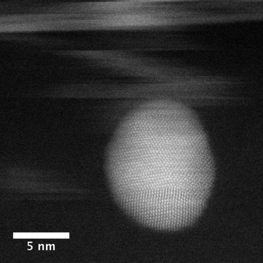 High-resolution
Z-contrast image of a gold nanoparticle in a liquid
(hexane) imaged using fluid cell STEM. The blurred areas are caused by CdSe
nanoparticle rapidly moving in the liquid during the recording of the image.
|
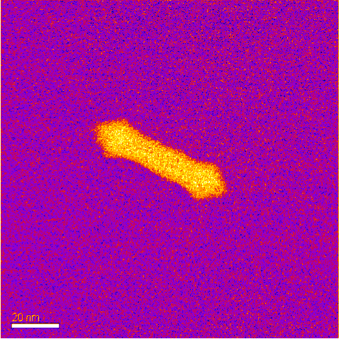 Color-coded
Z-contrast image of a gold nanorod in water after precipitation of
calcite from amorphous calcium carbonate. Calcite nanocrystals have
formed at the tips of the nanrod.
|
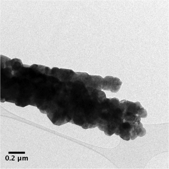 Bright-field
TEM image of aragonite precipitated from a supersaturated solution of
calcium and carbonate ions in the presence of ethanol.
|
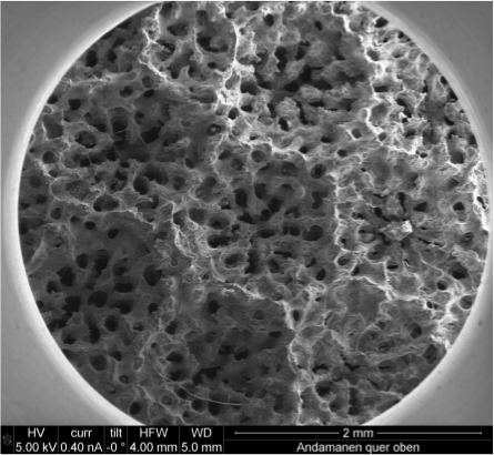
Scanning electron
micrograph of the top of the skeleton of the coral Porites lobata.
|
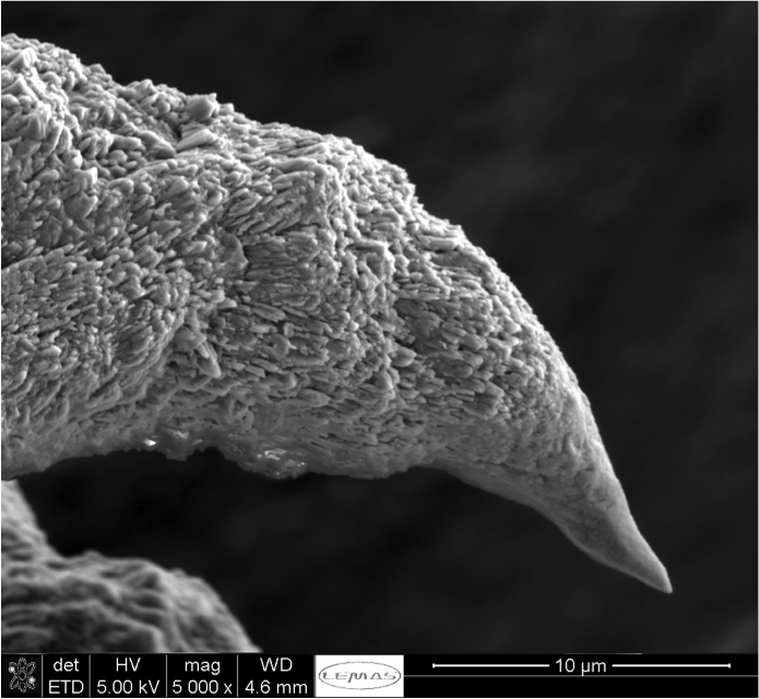 A spicule formed
during the juvenile stage of coral growth imaged by SEM. A spicule formed
during the juvenile stage of coral growth imaged by SEM. |
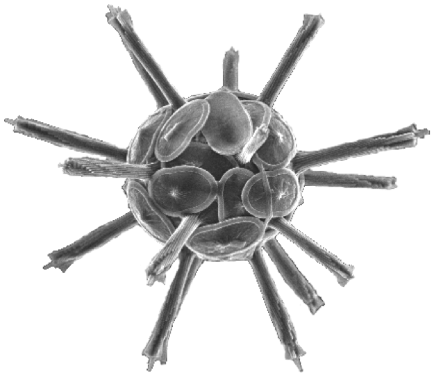 A coccolithophore of
the calcifying algae Rhabdosphaera clavigera imaged by SEM.
|
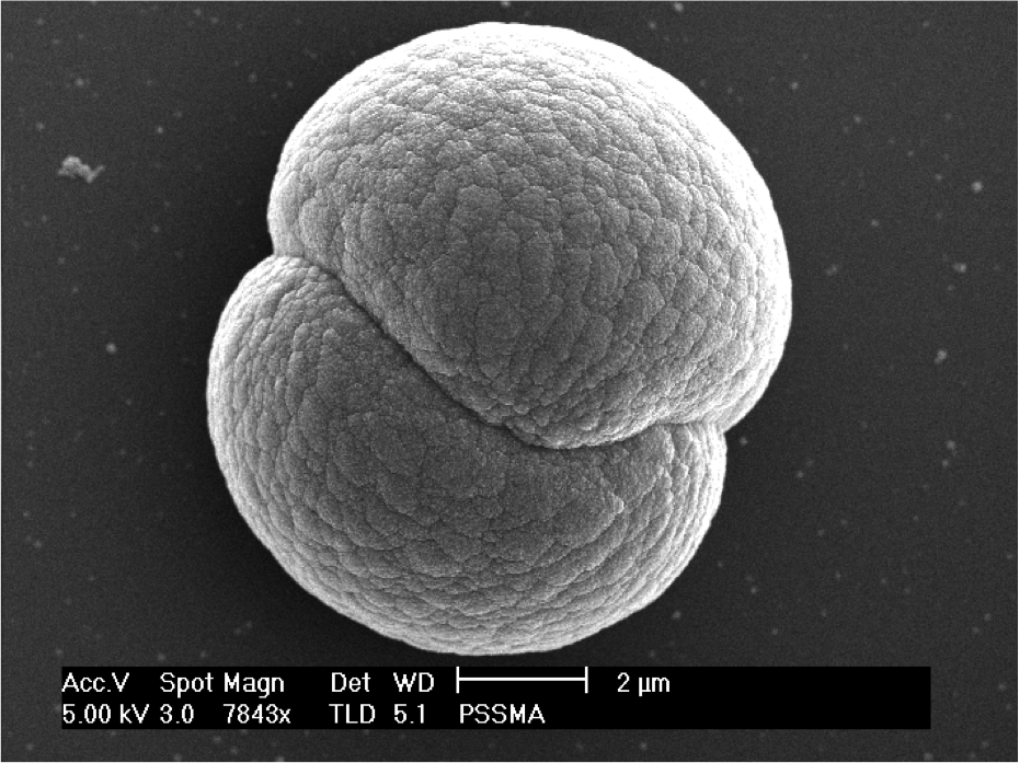 Calcium carbonate
precipiated in the presence of the polymer PMMA imaged by SEM.
|
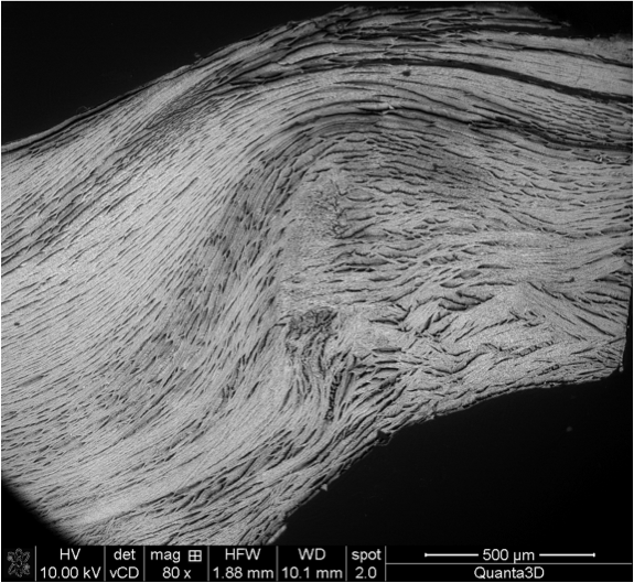 Cross-sectional view
of the scallop Pekten maximus
after mild etching to visualise the layered structure formed by
calcite nanocrystals.
|
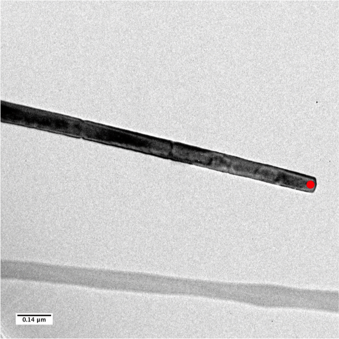 Single
crystalline calcite nanowire deposited using a track-etch membrane.
|