Kystis Ex Vivo Human Bladder Research - The University of York
How can we help?
Call us on +44 (0) 1904 328766 or
email us at info@kystis.com

Our assays
Kystis is continuously working with customers to develop project-specific assays and the examples below have all been previously validated as moderate (96-well) throughput assays:
Functional differentiation is the key to the added value of the Biomimetic Urothelium over existing proliferative cell models. Several different strategies have been developed to look at urothelial differentiation within this tissue model.
- Differentiation-associated protein expression can be monitored in 96-well plates using Li-Cor In cell Westerns to label for example Cytokeratins or Claudins.
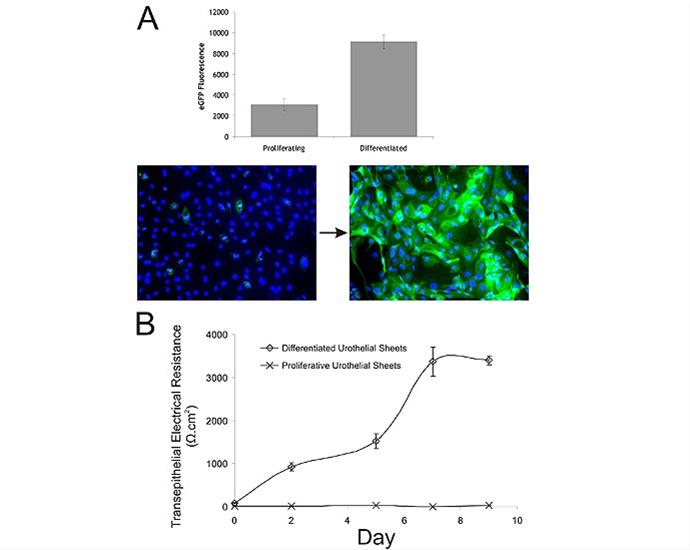
- The uroplakins are a family of barrier proteins only expressed by the urothelium. We have made a uroplakin II lentiviral reporter (which links the promoter of uroplakin II with GFP expression) to assess the status of urothelial differentiation in real-time (Figure A).
- Transepithelial electrical resistance monitoring (TER) is a physical assessment how “tight” the barrier is and again can be monitored in real-time. Figure B shows the formation of a strong barrier over a 9 day timecourse.
- ERK, AKT, WNT protein expression (total and active forms)
Human urothelial tissues are naturally highly quiescent with very little cell division. This is one of the reasons proliferative cell cultures are poorly representative of the native tissue. The figure below shows the Alamar Blue assay used to illustrate the reduced cell division within the differentiated Biomimetic Urothelium.

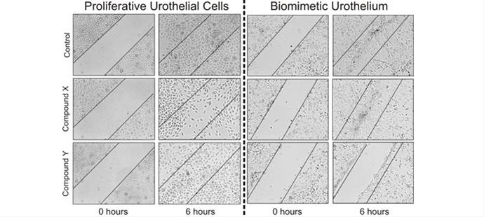
Scratch wound healing assays have proved very informative. When conducted over short periods of time, induced proliferation can play no role and this allows easily quantifiable study of migration. The figure below shows how two compounds affect urothelial wound healing in opposite fashions depending on the differentiation state of the tissue. These results are from unpublished data and compounds will be revealed following publication.
Apoptosis can be monitored by any of the wide range of existing assays. We have characterised Alamar Blue, Live/Dead staining and an active caspase 3/7 assay for this purpose.
- Affymetrix arrays, quantitative realtime PCR and next generation sequencing
- shRNA, siRNA technologies to knock down target gene expression retrovirus technologies to overexpress wildtype, oncogenic or dominant-negative genes